


 Digital Radiography
Dental X-rays are a useful diagnostic tool when helping your dentist detect damage and disease not visible during a regular dental exam. How often X-rays should be taken depends on your present oral health, your age, your risk for disease, and any signs and symptoms of oral disease. For example, children may require X-rays more often than adults because their teeth and jaws are still developing and their teeth are more likely to be affected by tooth decay than those of adults. Your dentist will review your history, examine your mouth and then decide whether or not you need X-rays.
If you are a new patient, the dentist may recommend X-rays to determine the present status of your oral health and have a baseline to help identify changes that may occur later. A new set of X-rays may be needed to help your dentist detect any new cavities, determine the status of your gum health or evaluate the growth and development of your teeth. If a previous dentist has any radiographs of you, your new dentist may ask you for copies of them. Ask both dentists to help you with forwarding your X-rays.
Dental X-ray exams are safe; however, they do require very low levels of radiation exposure, which makes the risk of potentially harmful effects very small. Dental X-ray tools and techniques are designed to limit the body's exposure to radiation and every precaution is taken to ensure that radiation exposure is As Low As Reasonable Achievable (the ALARA principle). A leaded apron minimizes exposure to the abdomen and may be used when it will not interfere with acquisition of the dental radiograph. Also, a leaded thyroid collar can protect the thyroid from radiation, and should also be used whenever possible. The use of a leaded thyroid collar is recommended for women of childbearing age, pregnant women and children.
If you are pregnant, tell your dentist. During your pregnancy, you may need to have X-rays taken as part of your treatment plan for a dental disease. Use of the leaded apron and thyroid collar will protect you and your fetus from radiation exposure. Dental X-rays do not need to be delayed if you are trying to become pregnant or are breastfeeding.
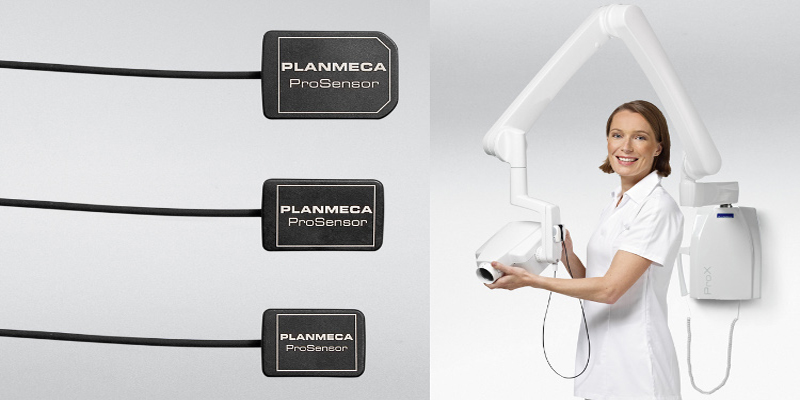
We have the best imagiology methods, allowing to raise the quality and quickness of diagnosis and enabling the treatment planning in an accurately and predictable way.There are two main types of dental X-rays: intraoral (meaning the X-ray film is inside the mouth) and extraoral (meaning the X-ray film is outside the mouth).
- Intraoral X-rays are the most common type of dental X-ray taken. These X-rays provide a lot of detail and allow your dentist to find cavities, check the health of the tooth root and bone surrounding the tooth, check the status of developing teeth, and monitor the general health of your teeth and jawbone.
- Extraoral X-rays show teeth, but their main focus is the jaw and skull. These X-rays do not provide the detail found with intraoral X-rays and therefore are not used for detecting cavities or for identifying problems with individual teeth. Instead, extraoral X-rays are used to look for impacted teeth, monitor growth and development of the jaws in relation to the teeth, and to identify potential problems between teeth and jaws and the temporomandibular joint (TMJ) or other bones of the face.
Types of Intraoral X-Rays
There are several types of intraoral X-rays, each of which shows different aspects of teeth.
- Bite-wing X-rays show details of the upper and lower teeth in one area of the mouth. Each bite-wing shows a tooth from its crown to about the level of the supporting bone. Bite-wing X-rays are used to detect decay between teeth and changes in bone density caused by gum disease. They are also useful in determining the proper fit of a crown (or cast restoration) and the marginal integrity of fillings.
- Periapical X-rays show the whole tooth - from the crown to beyond the end of the root to where the tooth is anchored in the jaw. Each periapical X-ray shows this full tooth dimension and includes all the teeth in one portion of either the upper or lower jaw. Periapical X-rays are used to detect any abnormalities of the root structure and surrounding bone structure.
- Occlusal X-rays are larger and show full tooth development and placement. Each X-ray reveals the entire arch of teeth in either the upper or lower jaw.
Types of Extraoral X-Rays
There are several types of extraoral X-rays that your dentist may take.
- Panoramic X-rays show the entire mouth area -- all the teeth in both the upper and lower jaws -- on a single X-ray. This type of X-ray is useful for detecting the position of fully emerged as well as emerging teeth, can identify impacted teeth, and aid in the diagnosis of tumors.
- Tomograms show a particular layer or "slice" of the mouth while blurring out all other layers. This type of X-ray is useful for examining structures that are difficult to clearly see -- for instance, because other structures are in very close proximity to the structure to be viewed.
- Cephalometric projections show the entire side of the head. This type of X-ray is useful for examining the teeth in relation to the jaw and profile of the individual. Orthodontists use this type of X-ray to develop their treatment plans.
- Computed tomography, otherwise known as CT scanning, shows the body's interior structures as a three-dimensional image. This type of X-ray, which may be performed in a hospital or radiology center or a dental office, is used to identify problems in the bones of the face, such as tumors or fractures. CT scans are also used to evaluate bone for the placement of dental implants and difficult extractions. This helps the surgeon avoid possible complications during and after a surgical procedure.
![]()